Conditions
- Myopia
- Astigmatism
- Hyperopia
- Presbyopia
- Cataract
- Secondary Cataract
- Keratoconus
- Age-related Macular Degeneration
- Diabetic Retinopathy
- Glaucoma
- Dry Eye
- “Floaters” and Flashes
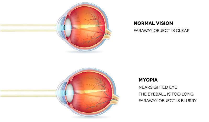
Myopia (nearsightedness) is a refractive error. Refractive error is when the eye does not bend (refract) light properly. Light does not focus correctly so images are not clear. In myopia, close objects look clear but distant objects appear blurred. Myopia is a common condition that affects an estimated 25% of Americans. It is an eye focusing disorder, not an eye disease. The eye’s tear film, cornea and lens bend light so it focuses on the retina. The retina receives the picture formed by these light rays. It sends the picture to the brain through the optic nerve, which is actually part of the brain.
Myopia occurs when the eye is longer than normal or has a cornea that is too steep. As a result, light rays focus in front of the retina instead of on it. In this case, you see near objects clearly, but distant objects will appear blurred.
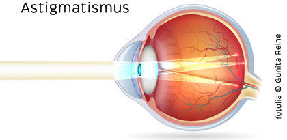
Astigmatism is an imperfection in the curvature of your eye’s cornea or lens. Normally, the cornea and lens are smooth and curved equally in all directions. This helps to focus light rays sharply onto the retina at the back of your eye. If your cornea or lens isn’t smooth and evenly curved, light rays aren’t refracted (bent) properly. Doctors call this a refractive error. An eye with all major parts of anatomy labeledWhen your cornea has an irregular shape, you have corneal astigmatism. When the shape of your lens is distorted, you have lenticular astigmatism. In either case, your vision for both near and far objects is blurry or distorted. It’s almost like looking into a fun house mirror in which you can appear too tall, too short, too wide or too thin. People may have astigmatism along with other refractive errors. Those errors may include things like: nearsightedness (myopia) or farsightedness (hyperopia). Adults with significant astigmatism may realize their vision isn’t as good as it should be. Children with astigmatism symptoms may not be aware they have this condition. They are unlikely to complain about blurred or distorted vision. An eye that focuses light correctly onto the retina In a normal eye, the cornea and lens focus light rays on the retina. An eye with astigmatism focuses light incorrectly. Light is focused both in front of and behind the retina. In astigmatism, images focus in front of and beyond the retina. Close and distant objects both appear blurry. Uncorrected astigmatism can impact a child’s ability to achieve in school and sports. It is crucial that children have regular eye exams. Get these exams to detect astigmatism and other vision problems as early as possible. What Causes Astigmatism? Astigmatism is caused by an irregular curvature of the eye’s cornea or lens. If your cornea or lens isn’t evenly curved, light rays aren’t refracted properly. With astigmatism you have blurred or distorted vision at near and far distances. Astigmatism is very common. Doctors don’t know why corneal shape differs from person to person. They do know that likelihood of developing astigmatism is inherited. Astigmatism can develop after an eye disease, eye injury or surgery. It is a myth that astigmatism can develop or worsen from reading in low light or sitting very close to the television. Astigmatism Symptoms Astigmatism symptoms may include: blurry vision or areas of distorted vision eyestrain headaches squinting to try to see clearly, or eye discomfort If you have these symptoms you may not necessarily have astigmatism. You should visit to your ophthalmologist. A complete eye exam will determine what is causing your symptoms.
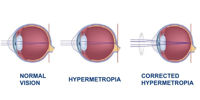
What is hyperopia?
Hyperopia is a condition in which light is focused behind the retina instead of on it, causing objects to be blurry.
In hyperopia, patients usually cannot see objects in close distances well, but they can see far objects normally, even though in some cases far sight may also be affected.
Hyperopia causes include heredity, axial length of the eyeball and reduced refractive power of the eye.
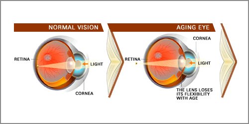
Presbyopia is a condition associated with the aging of the eye which results in eyes losing the ability to focus clearly on close objects.
It is a natural condition which affects almost everyone after the age of 40 to 50.
Presbyopia has been the subject of study for the international ophthalmic community. It is a very complex condition involving structures of both the brain and the eye.
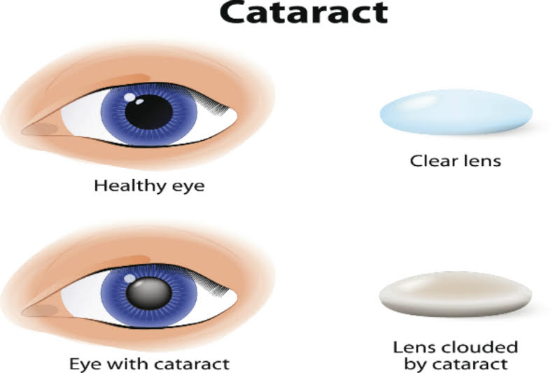
Inside our eyes, we have a natural lens. The lens bends (refracts) light rays that come into the eye to help us see. The lens should be clear, like the top lens in the illustration.
Vision Problems with Cataracts:
If you have a cataract, your lens has become cloudy, like the bottom lens in the illustration. It is like looking through a foggy or dusty car windshield. Things look blurry, hazy or less colorful with a cataract. The definition of a cataract is a cloudy lens in the eye, whatever the cause may be. Here the cataract lens is compared to a natural clear lens. The top lens is a clear, natural lens. The bottom lens shows clouding by cataract.
What Are the Symptoms of Cataracts?
Here are some vision changes you may notice if you have a cataract:
Having blurry vision.
Seeing double (when you see two images instead of one).
Being extra sensitive to light.
Having trouble seeing well at night, or needing more light when you read.
Seeing bright colors as faded or yellow instead.
Dull or yellowed vision from cataracts.
Dull or yellow vision from cataracts.
Blurry or dim vision from cataracts.
Blurry or dim vision is a symptom of cataracts.
Distortion or doubled images from cataracts.
Distortion or ghost images from cataracts.
See a simulation of what vision with cataract looks like. What Causes Cataracts?
Aging is the most common cause. This is due to normal eye changes that happen starting around age 40. That is when normal proteins in the lens start to break down. This is what causes the lens to get cloudy. People over age 60 usually start to have some clouding of their lenses. However, vision problems may not happen until years later.
Other reasons you may get cataracts include:
having parents, brothers, sisters, or other family members who have cataracts.
having certain medical problems, such as diabetes.
having had an eye injury, eye surgery, or radiation treatments on your upper body.
having spent a lot of time in the sun, especially without sunglasses that protect your eyes from damaging ultraviolet (UV) rays.
using certain medications such as corticosteroids, which may cause early formation of cataracts.
Most age-related cataracts develop gradually. Other cataracts can develop more quickly, such as those in younger people or those in people with diabetes. Doctors cannot predict how quickly a person’s cataract will develop.
You may be able to slow down your development of cataracts.
Protecting your eyes from sunlight is the best way to do this. Wear sunglasses that screen out the sun’s ultraviolet (UV) light rays. You may also wear regular eyeglasses that have a clear, anti-UV coating. Talk with your eye doctor to learn more.
Posterior Capsule Opacity
In a cataract operation, the cloudy lens is replaced with a clear, artificial lens. However, in some cases, a hazy membrane can form just behind the intraocular lens implant. This is known as opacity of the posterior capsule, or secondary cataract.
Symptoms A posterior capsule opacity will only occur after cataract surgery. If you have had a cataract operation, and you have blurred, hazy vision, or see a lot of glare from lights, it may be because of a posterior capsule opacity. Blurring and loss of vision from posterior capsule opacity is usually gradual, just as with real cataracts. Treatment The treatment for posterior capsule opacity is very simple. A procedure called a YAG laser capsulotomy is used to remove the haziness, and restore normal vision. It is a fast, painless and very effective treatment. It only takes 5 minutes and can be done in our center.
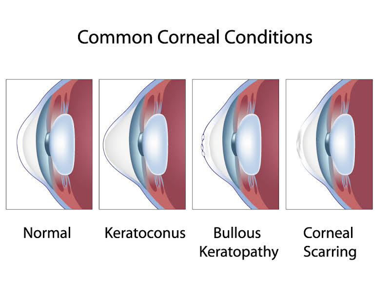
Keratoconus is a vision disorder that occurs when the normally round cornea (the front part of the eye) becomes thin and irregular (cone) shaped. This abnormal shape prevents the light entering the eye from being focused correctly on the retina and causes distortion of vision. In its earliest stages, keratoconus causes slight blurring and distortion of vision and increased sensitivity to glare and light. These symptoms usually appear in the late teens or late 20s. Keratoconus may progress for 10-20 years and then slow in its progression. Each eye may be affected differently. As keratoconus progresses, the cornea bulges more and vision may become more distorted. In a small number of cases, the cornea will swell and cause a sudden and significant decrease in vision. The swelling occurs when the strain of the cornea’s protruding cone-like shape causes a tiny crack to develop. The swelling may last for weeks or months as the crack heals and is gradually replaced by scar tissue. If this sudden swelling does occur, your doctor can prescribe eyedrops for temporary relief, but there are no medicines that can prevent the disorder from progressing. The right diagnosis and the exact follow can only be done with CORNEAL TOMOGRAPHY. Our center is equipt with the most modern Tomography from the German firma Oculus, Pentacam AXL.
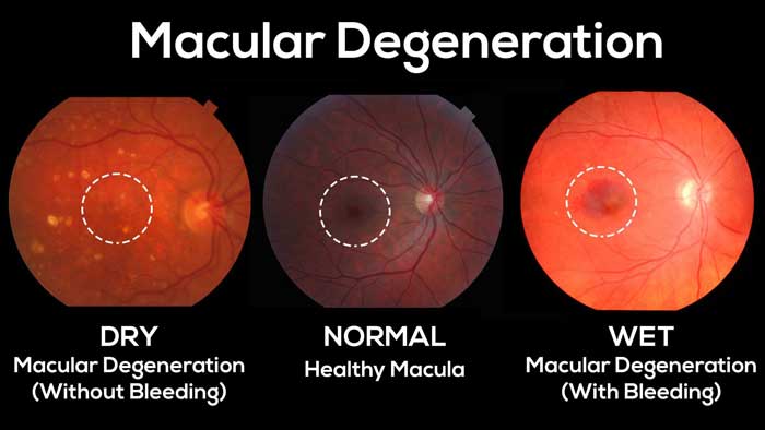
Age-related Macular Degeneration (AMD) is one of the main causes of eyesight loss in people over 60.
There are two types of Age-related Macular Degeneration Dry and
Wet (exudative).
The dry type affects approximately 85% of individuals with AMD. Photo-receptors in the center of the retina are destroyed. This leads to a decrease of central vision, which affects everyday activities, such as reading and driving. Dry type AMD can evolve to wet type AMD.
In wet (or exudative) type AMD, abnormal vessels develop beneath the retina, in the choroid of the macula. These may cause edema, hemorrhage and finally a scar on the macula. This causes a significant loss of central vision, central scotoma or metamorfopsia (straight lines appear wavy). As the macula is at the center of the retina, in macular degeneration peripheral vision remains unchanged.
The right diagnosis and treatment should take early enough place. Do not hecitate to contact us.
People with diabetes can have an eye disease called diabetic retinopathy. This is when high blood sugar levels cause damage to blood vessels in the retina. These blood vessels can swell and leak. Or they can close, stopping blood from passing through. Sometimes abnormal new blood vessels grow on the retina. All of these changes can steal your vision.
Stages of Diabetic Eye Disease:
There are two main stages of diabetic eye disease.
NPDR (non-proliferative diabetic retinopathy)
This is the early stage of diabetic eye disease. Many people with diabetes have it.
With NPDR, tiny blood vessels leak, making the retina swell. When the macula swells, it is called macular edema. This is the most common reason why people with diabetes lose their vision.
Also with NPDR, blood vessels in the retina can close off. This is called macular ischemia. When that happens, blood cannot reach the macula. Sometimes tiny particles called exudates can form in the retina. These can affect your vision too.
If you have NPDR, your vision will be blurry.
PDR (proliferative diabetic retinopathy)
PDR is the more advanced stage of diabetic eye disease. It happens when the retina starts growing new blood vessels. This is called neovascularization. These fragile new vessels often bleed into the vitreous. If they only bleed a little, you might see a few dark floaters. If they bleed a lot, it might block all vision.
These new blood vessels can form scar tissue. Scar tissue can cause problems with the macula or lead to a detached retina.
PDR is very serious, and can steal both your central and peripheral (side) vision.
Diabetic Retinopathy Symptoms
You can have diabetic retinopathy and not know it. This is because it often has no symptoms in its early stages. As diabetic retinopathy gets worse, you will notice symptoms such as:
- seeing an increasing number of floaters,
- having blurry vision,
- having vision that changes sometimes from blurry to clear,
- seeing blank or dark areas in your field of vision,
- having poor night vision
- noticing colors appear faded or washed out losing vision.
Diabetic retinopathy symptoms usually affect both eyes.
Glaucoma is a disease that damages your eye’s optic nerve. It usually happens when fluid builds up in the front part of your eye. That extra fluid increases the pressure in your eye, damaging the optic nerve.
Types of glaucoma
The types of glaucoma are:
Open-angle glaucoma
Acute and chronic angle-closure glaucoma
Pseudoexfoliative glaucoma
Pigmentary glaucoma
Normal tension glaucoma
Congenital glaucoma
Proper examinations and tests should take place , in the early stages.

Our eyes need tears to stay healthy and comfortable. If your eyes do not produce enough tears, it is called dry eye. Dry eye is also when your eyes do not make the right type of tears or tear film.
How Do Tears Work?
When you blink, a film of tears spreads over the eye. This keeps the eye’s surface smooth and clear. The tear film is important for good vision.
The tear film is made of three layers:
An oily layer A watery layer A mucus layer
Each layer of the tear film serves a purpose.
The oily layer is the outside of the tear film. It makes the tear surface smooth and keeps tears from drying up too quickly. This layer is made in the eye’s meibomian glands.
The watery layer is the middle of the tear film. It makes up most of what we see as tears. This layer cleans the eye, washing away particles that do not belong in the eye. This layer comes from the lacrimal glands in the eyelids.
The mucus layer is the inner layer of the tear film. This helps spread the watery layer over the eye’s surface, keeping it moist. Without mucus, tears would not stick to the eye. Mucus is made in the conjunctiva. This is the clear tissue covering the white of your eye and inside your eyelids.
Normally, our eyes constantly make tears to stay moist. If our eyes are irritated, or we cry, our eyes make a lot of tears. But, sometimes the eyes don’t make enough tears or something affects one or more layers of the tear film. In those cases, we end up with dry eyes.
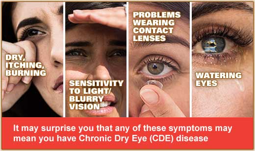
Dry Eye Symptoms
Here are some of the symptoms of dry eye.
You feel like your eyes are stinging and burning. There is a scratchy or gritty feeling like something is in your eye. There are strings of mucus in or around your eyes. Your eyes are red or irritated. This is especially true when you are in the wind or near cigarette smoke. It is painful to wear contact lenses. You have lots of tears in your eyes.
Having a lot of tears in your eyes with “dry eye” might sound odd. But your eyes make more tears when they are irritated by dry eye. Dry Eye Causes
People tend to make fewer tears as they get older due to hormonal changes. Both men and women can get dry eye. However, it is more common in women—especially those who have gone through menopause.
Here are some other causes of dry eye.
Certain diseases, such as rheumatoid arthritis, Sjögren’s syndrome, thyroid disease, and lupus Blepharitis (when eyelids are swollen or red) Entropion (when eyelids turn in); ectropion (eyelids turn outward) Being in smoke, wind or a very dry climate Looking at a computer screen for a long time, reading and other activities that reduce blinking Using contact lenses for a long time Having refractive eye surgery, such as LASIK Taking certain medicines, such as: Diuretics (water pills) for high blood pressure Beta-blockers, for heart problems or high blood pressure Allergy and cold medicines (antihistamines) Sleeping pills Anxiety and antidepressant medicines Heartburn medicines
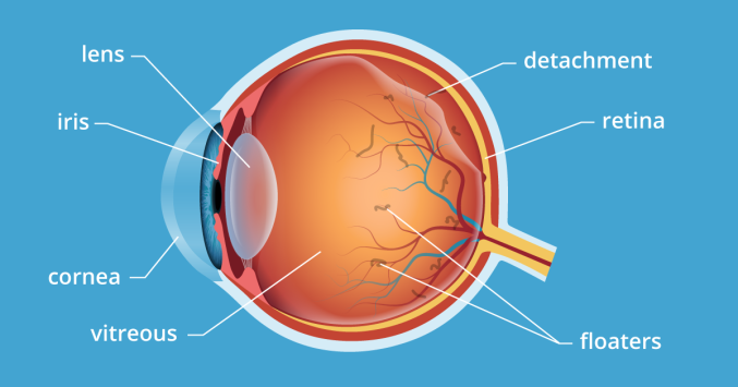
Floaters look like small specks, dots, circles, lines or cobwebs in your field of vision. While they seem to be in front of your eye, they are floating inside. Floaters are tiny clumps of gel or cells inside the vitreous that fills your eye. What you see are the shadows these clumps cast on your retina. You usually notice floaters when looking at something plain, like a blank wall or a blue sky. As we age, our vitreous starts to thicken or shrink. Sometimes clumps or strands form in the vitreous. If the vitreous pulls away from the back of the eye, it is called posterior vitreous detachment. Floaters usually happen with posterior vitreous detachment. They are not serious, and they tend to fade or go away over time. Severe floaters can be removed by surgery, but this is seldom necessary. You are more likely to get floaters if you: are nearsighted (you need glasses to see far away) have had surgery for cataracts have had inflammation (swelling) inside the eye What are flashes? Flashes can look like flashing lights or lightning streaks in your field of vision. Some people compare them to seeing “stars” after being hit on the head. You might see flashes on and off for weeks, or even months. Flashes happen when the vitreous rubs or pulls on your retina. As people age, it is common to see flashes occasionally. Flashes and migraines Sometimes people have light flashes that look like jagged lines or heat waves. These can appear in one or both eyes and may last up to 20 minutes. This type of flash may be caused by a migraine. A migraine is a spasm of blood vessels in the brain. When you get a headache after these flashes, it is called a “migraine headache.” But sometimes you only see the light flash without having a headache. This is called an “ophthalmic migraine” or “migraine without headache.”